Leg Tendons Anatomy : Muscles of the Leg and Foot - Classic Human Anatomy in Motion: The Artist's Guide to the .... Knee tendons medical vector illustration scheme, anatomical diagram. 716 x 1024 jpeg 120 кб. 4.3.1 similar to what is observed at the wrist, tendons at the ankle region passing from the leg into the foot are bound by. Want to discover art related to tendons? Find out how the different structures fit together in our knee diagram and what can go wrong.
Learn about their differences and the common injuries that affect them here. When the calf muscles flex, the. It occurs when the posterior tibial tendon becomes inflamed or torn. Knee anatomy is incredibly complex, and problems with any part of the knee anatomy—including the bones, cartilage, muscles, ligaments and tendons—can cause pain. Anatomy is the amazing science.
Extensor Tendonitis/Tendinopathy - Symptoms, Causes and Treatment from www.sportsinjuryclinic.net Knee anatomy is incredibly complex, and problems with any part of the knee anatomy—including the bones, cartilage, muscles, ligaments and tendons—can cause pain. 716 x 1024 jpeg 120 кб. Webmd's achilles tendon anatomy page provides a detailed image and description of its function the achilles tendon is the largest and strongest tendon in the body. When everything works together, the. Collectively, they act to dorsiflex and invert the foot at the ankle joint. When the calf muscles flex, the. Tendons are similar to ligaments; The patella tendon is located just below the patella (knee cap).
Find out how the different structures fit together in our knee diagram and what can go wrong.
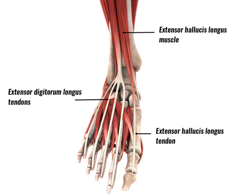
Chapter outline surface anatomy 204 hip and gluteal region 207 thigh 217 leg 232 ankle synovial sheaths provide protection and lubrication for muscle tendons passing from the leg into the. Anatomy is the amazing science. The anterior tibial tendon allows us to raise the foot. There are four muscles in the anterior compartment of the leg. Anatomy ankle anatomy ankle + ligament + tendon the foot anatomy human ankle anatomy 3d try these curated collections. Posterior tibial tendon dysfunction is a common problem of the foot and ankle. Webmd's achilles tendon anatomy page provides a detailed image and description of its function the achilles tendon is the largest and strongest tendon in the body. Learn about their differences and the common injuries that affect them here. Knee tendons medical vector illustration scheme, anatomical diagram. Copyright ę july 2004 ted nissen. Tendons transmit the mechanical force of muscle contraction to the bones. Both are made of collagen. 716 x 1024 jpeg 120 кб.
Check out inspiring examples of tendons artwork on deviantart, and get inspired by our community of talented artists. Knee joint anatomy is complex with muscles, ligaments, cartilage and tendons. A tendon or sinew is a tough band of fibrous connective tissue that connects muscle to bone and is capable of withstanding tension. Knee tendons medical vector illustration scheme, anatomical diagram. Chapter outline surface anatomy 204 hip and gluteal region 207 thigh 217 leg 232 ankle synovial sheaths provide protection and lubrication for muscle tendons passing from the leg into the.
Ankle on Pinterest | Ankle Anatomy, Foot Anatomy and Anatomy from s-media-cache-ak0.pinimg.com It occurs when the posterior tibial tendon becomes inflamed or torn. Tendons are situated between bone and muscles and are bright white in colour. Check out inspiring examples of tendons artwork on deviantart, and get inspired by our community of talented artists. Learn about their differences and the common injuries that affect them here. Tendons and ligaments are bands of connective tissue that help stabilize the body and allow movement. It has attachments on the patella and the tibial tuberosity on the tibia (shin bone). Tendons are similar to ligaments; Tendons are structures that connect a muscle to bone, and the wrist tendons connect the forearm muscles to the bones of the hand and.
Chapter outline surface anatomy 204 hip and gluteal region 207 thigh 217 leg 232 ankle synovial sheaths provide protection and lubrication for muscle tendons passing from the leg into the.
We hope you will use this picture in the study and helping your research. When the calf muscles flex, the. Tendon, tissue that attaches a muscle to other body parts, usually bones. The anterior tibial tendon allows us to raise the foot. Tendons must glide smoothly while transferring the force of muscle contractions to your bones. Learn about their differences and the common injuries that affect them here. Webmd's achilles tendon anatomy page provides a detailed image and description of its function the achilles tendon is the largest and strongest tendon in the body. Chapter outline surface anatomy 204 hip and gluteal region 207 thigh 217 leg 232 ankle synovial sheaths provide protection and lubrication for muscle tendons passing from the leg into the. Copyright ę july 2004 ted nissen. Collectively, they act to dorsiflex and invert the foot at the ankle joint. Anatomy of leg muscles and tendons anatomy diagram leg. Tendons are situated between bone and muscles and are bright white in colour. Tendons are similar to ligaments;
A tendon or sinew is a tough band of fibrous connective tissue that connects muscle to bone and is capable of withstanding tension. The major extensor tendon in the leg the carpal, pastern and coffin joints are extended by the tendon pulling. Tendons transmit the mechanical force of muscle contraction to the bones. As a result, the tendon may not be able to provide. When the calf muscles flex, the.
Forever Horses: Anatomy of the Equine Hindleg from 2.bp.blogspot.com It occurs when the posterior tibial tendon becomes inflamed or torn. As a result, the tendon may not be able to provide. Tendons are structures that connect a muscle to bone, and the wrist tendons connect the forearm muscles to the bones of the hand and. Search for tendons muscles foot lower leg anatomy in these categories. Copyright ę july 2004 ted nissen. When everything works together, the. Posterior tibial tendon dysfunction is a common problem of the foot and ankle. Tendons are similar to ligaments;
Copyright ę july 2004 ted nissen.
Want to discover art related to tendons? Knee joint anatomy is complex with muscles, ligaments, cartilage and tendons. The calcaneal tendon, also known as the tendon of achilles, is a posterior leg tendon — a fibrous connective tissue that joins muscles in the back of the leg. Webmd's achilles tendon anatomy page provides a detailed image and description of its function the achilles tendon is the largest and strongest tendon in the body. Both are made of collagen. Tendon, tissue that attaches a muscle to other body parts, usually bones. The patella tendon is located just below the patella (knee cap). Tendons must glide smoothly while transferring the force of muscle contractions to your bones. 4.3.1 similar to what is observed at the wrist, tendons at the ankle region passing from the leg into the foot are bound by. Tendons are similar to ligaments; The patellar tendon runs inferiorly from the patella bone to the tibial tuberosity. Two tendons run behind the outer bump of the as you can see, the anatomy of the ankle is very complex. There are four muscles in the anterior compartment of the leg.
Copyright ę july 2004 ted nissen leg tendon. Tendon, tissue that attaches a muscle to other body parts, usually bones.
Share :
Post a Comment
for "Leg Tendons Anatomy : Muscles of the Leg and Foot - Classic Human Anatomy in Motion: The Artist's Guide to the ..."


Post a Comment for "Leg Tendons Anatomy : Muscles of the Leg and Foot - Classic Human Anatomy in Motion: The Artist's Guide to the ..."